- Researchers have come up with a new method to measure brain’s electrical activity, without electrodes.
- They built Archon1, a light-sensitive protein, that ejects a fluorescent signal after it’s delivered into a brain cell.
Neurons communicate with each other through electrical events known as “action potentials” and chemical neurotransmitters. They enable the brain to harmonize thoughts, sensation, emotions and behavior.
Usually, studying this electrical activity involves measuring these impulses with electrodes embedded into the brain, which is a time-consuming and difficult task. Also, it’s not much efficient.
Researchers at MIT have discovered a new method to measure brain’s electrical activity, and they claim that it’s more informative and much easier. They’ve built a special protein sensitive to light, which could be inserted into neuron membranes, where it ejects a fluorescent signal, indicating the amount of voltage a specific cell is experiencing.
This study could allow researchers to better understand how neurons behave, as the brain executes a specific function. Neural activity of several cells can now be recorded and monitored, millisecond by millisecond.
Creating Fluorescent-Sensitive Protein
Developing fluorescent-sensitive molecules was not an easy task at all. It requires protein to rapidly respond and doesn’t fade when exposed to light (or in other words, should be photobleaching resistant).
To develop such molecule, researchers created a small robot for screening million of proteins via ‘directed protein evolution’. The idea is to make millions of mutant genes from a single gene, and finally pick a few of them that function the best (as per the requirement).
They created one and a half million mutated protein (sensitive to light) and named them QuasAr2. Then they put all of these proteins into mammalian cells (1 mutant in each cell). Researchers let these cells grow in lab equipments and pictured them using an advanced microscope.
 Image credit: UCI Applied Innovation
The custom-built robot then identified cells with proteins that met the researchers’ requirement. These requirements primarily include the brightness level and the location of protein within the cell.
The next step was to select 5 best cells and perform one more mutation round, to produce another 8 million cells. The robot again identified the best 7 out of 8 million cells. Finally, researchers selected the 1 top performer out of these 7, which they named Archon1.
Reference: Nature Chemical Biology | doi:10.1038/s41589-018-0004-9 | MIT
Measuring Brain’s Electrical Activity Using Archon1
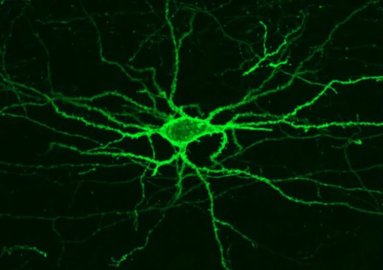 Representative image of Archon1 expressing neuron mouse motor cortex
Once Archon1 is injected into a cell, it automatically inserts itself within the cell membrane and provides a precise measurement of voltage of the cell. This allows scientists to calculate the electrical activity in brain cell of mouse, Caenorhabditis elegans worm and zebrafish larvae.
Archon1 emits a longer wavelength of red light, when the cells are subjected to a particular orangish-red light. The light’s brightness emitted by Archon1 indicates the particular cell’s voltage at a specific moment.
Archon1 could also be used with other light-sensitive molecules, which are typically used to stimulate or silence neuron activity as far as proteins react to any color except red. Such type of proteins is called optogenetic proteins.
Archon1 exhibits zero photobleaching under 8 minutes of continuous excitation with power similar to those used in the voltage imaging experiments, thus supporting neural activity recordings over behaviorally relevant timescales.
What’s Next?
The study opens up the new possibility for simultaneous recordings of large quantity of neurons with single-cell single-spike resolution in vivo. The existing scientific-grade cameras can perform fast imaging (from 500 to 1,000 Hz) over pixel counts smaller by an order of magnitude than those usually used for calcium imaging (from 10 to 20 Hz). New cameras with fast imaging capabilities at cellular resolution over broader fields of view will continue to improve the power of voltage imaging.
The research team plans to use this technique to calculate mice’s brain activity as they execute different tasks. This would make mapping neural circuits easier, and help researchers understand how they generate certain behaviors.
Read: 15 Mind-boggling Facts About The Nervous System
In coming years, researchers would be able to observe a neural computation taking place. They’ll be trying to resolve a few tiny brain circuits entirely, which could eventually help us understand what a feeling or a thought actually is.